
Figure 1.
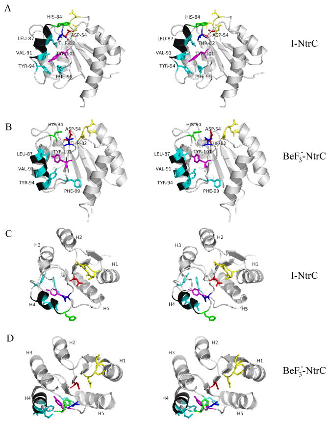
Stereoview of briefly minimized NMR structures of NtrCr. A) Side view of I-NtrCr, B) side view of , C) top view of I-NtrCr, D) top view of . The key helix, α4, is shown in black. Locations of residues Thr-82 (blue), Leu-87, Val-91, Tyr-94, Phe-99 (cyan), Tyr-101 (purple), Asp-54 (red), and His-84 (green) is indicated in top two figures. Residues Asp-10, Asp-11, Asp-12 are shown in yellow.