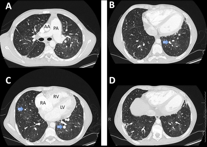
Figure 3.
Radiographic findings of pulmonary hypertension in an 18-year-old patient with sickle cell disease. (A) Note the central pulmonary artery (PA) is enlarged and significantly larger than the adjacent ascending aorta (AA), indicating a PA/AA ratio greater than 1. (B, C) Relative increase in segmental artery size relative to adjacent bronchus (arrows) as well as loss of peripheral vascularity. (C) Note right ventricle (RV) is hypertrophied and dilated with evidence of right atrial (RA) dilation. All images illustrate the finding of mosaic perfusion pattern of parenchymal attenuation.

