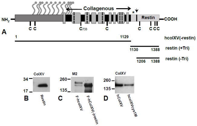
Figure 1. Diagram to show the structure of the collagen XV protein (A) and expression hcolXV domains and mutants (B–D).
A, (From (Amenta et al., 2005)). Amino terminal non-collagenous domain (dark gray); carboxyl domain (restin) (pale gray, with arrow marking location of cleavage sites); collagenous domain (gray boxes) with interruption marked as dashed lines or black bars depending on their length. Ball and stick symbols show consensus sites for GAG attachment; C denotes cysteines; * denotes trimerization domain; horizontal black bars denote the extent of the N-terminal and C-terminal domain constructs. B–D. Western blots of lysates from COS-7 cells transiently transfected with B) restin alone; C) F-hcolXV and F-hcolXV(−)restin; D) hcolXV and hcolXVCys1M). B) and D) probed with anti-colXV antibody and C) with M2 (anti-FLAG). MWs are shown in kDa.

