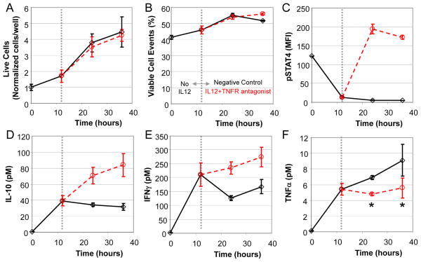
Fig. 4.
An autocrine feedback loop regulates TNFα expression in 2D6 cells that is independent of STAT4 activation. Following a 12 hour pre-conditioning with cRPMI, 2D6 cells were stimulated with IL-12 in combination with a TNF receptor antagonist (circles). Untreated cells were used as a negative control (diamonds). Changes in live cell density (A), viable cellular events (B), and STAT4 activation (C) were quantified as a function of time by flow cytometry. Enrichment of IL-10 (D), IFNγ (E), and TNFα (F) in the 2D6-conditioned media were quantified as a function of time using cytometric bead array. The dotted vertical line indicates the switch from pre-conditioning to stimulation conditions. Mean response (± standard deviation) at each time point and condition were used to create trend lines (solid and dotted lines). A Student’s t test was used to assess statistical significance, where * indicates p<0.05.

