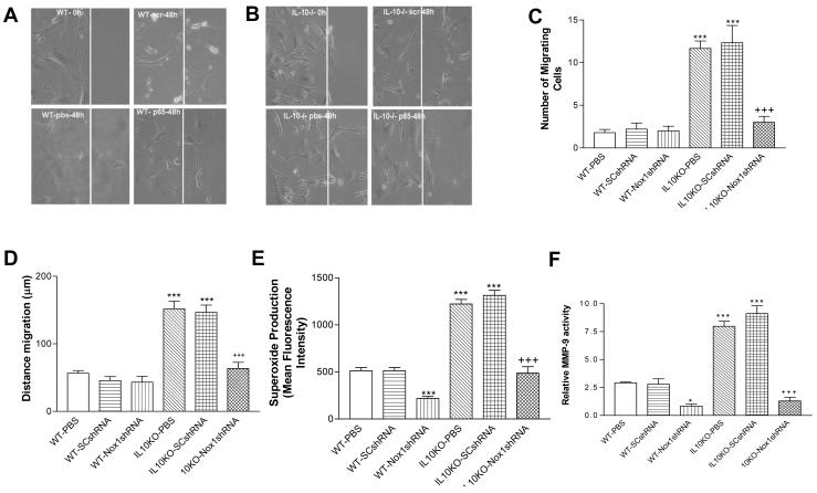
Figure 4. Effects of RNAi silencing of Nox1 on cell migration, ROS production and MMP-9 Activity in isolated and cultured smooth muscle cells (SMCs).
Confluent cells were allowed to grow for 48 h following wound injury. A photograph of cells migrated after 48 h in each group (A&B). Scr, scrambled shRNA. p65, Nox1shRNA. IL-10-/-, IL10 gene knockout. pbs, PBS (phosphate buffer solution). The summary data of the number of cells migrated and the average distance traveled (C&D). Aortic SMCs were exposed to the superoxide specific dye dihydroethidium (10 μM) for 30 min. Ethidium fluorescence was measured quantitatively by flow cytometry (E). MMP-9 activity was measured by gelatin zymography (F). Data=means±SE. *p<0.05, ***p<0.001 vs the WT-PBS group. +++p<0.001vs the IL10KO-PBS group. N=5.

