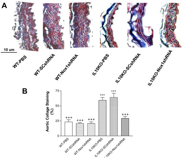
Figure 5. Effects of RNAi silencing of Nox1 on the aortic collagen content.
Trichrome staining of the aorta sections (7 μM) from IL-10KO and WT mice. Heavy collagen staining (blue) was found in the SMC layer and the adventitial matrix in the IL10KO mice (A). There was a loss of elastin fibers in the medial layer in the IL10KO mice (A). Quantification of collagen content (B). Data=means±SE. ***p<0.001 vs the WT-PBS group. +++p<0.001vs the IL10KO-PBS group. N=5.

