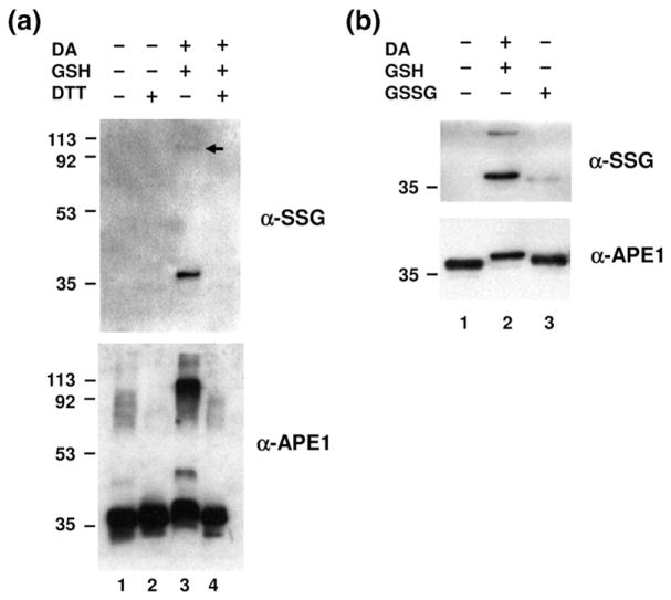
Fig. 1.
S-Glutathionylation of APE1 in vitro. (a) Purified recombinant APE1 protein (0.2 μg) was incubated with 5 mM diamide (DA) and 10 mM GSH as described in Materials and Methods. For deglutathionylation, S-glutathionylated APE1 was treated with 10 mM DTT. All samples were subjected to nonreducing SDS-PAGE followed by Western blot analysis with anti-SSG antibody (top) or anti-APE1 antibody (bottom). Positions of molecular mass protein standards are designated in kilodaltons, and the arrow denotes location of multi-meric protein form. The data are representative of at least three independent experiments. (b) S-Glutathionylation was carried out by disulfide exchange with GSSG. Recombinant APE1 protein (0.2 μg) was treated with either 5 mM diamide/10 mM GSH (lane 2) or 10 mM GSSG (lane 3), followed by nonreducing SDS-PAGE and Western blot analysis with anti-SSG antibody or anti-APE1 antibody. The data are representative of at least three independent experiments.

