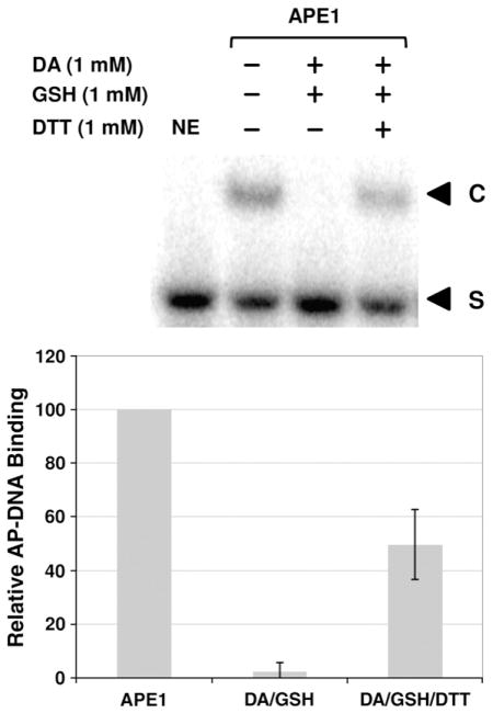
Fig. 3.
Effect of S-glutathionylation on APE1 DNA binding. After treatment with 1 mM diamide (DA), 1 mM GSH, and/or 1 mM DTT (as indicated), AP-site-containing DNA (100 fmol) was incubated with 2 ng of APE1 protein and then resolved on an 8% non-denaturing polyacrylamide gel (see Materials and Methods). (Top) A representative gel image of three independent experimental EMSAs. The bottom graph shows the relative DNA binding activity (compared to APE1 without DA, GSH, or DTT treatment) with means±SD (n=3). NE, no enzyme and DNA-only control; S, unbound substrate; C, APE1/AP–DNA complexes.

