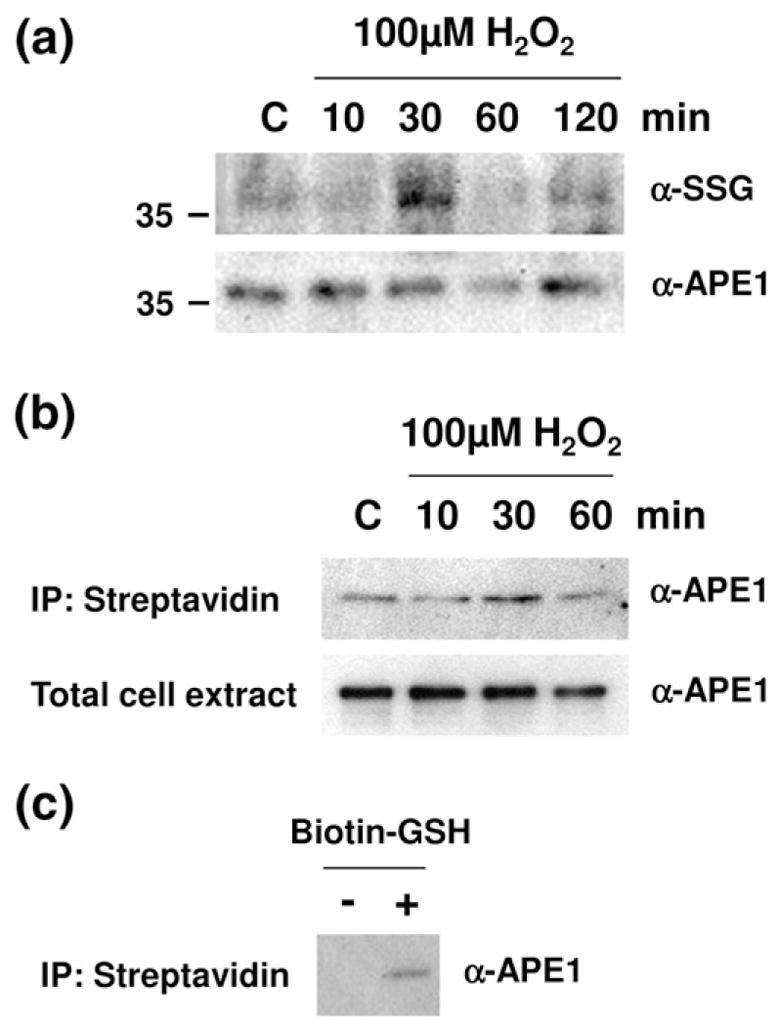
Fig. 6.

H2O2-induced S-glutathionylation of APE1 in cells of APE1 in cells (a) HeLa cells were exposed to 100 μM H2O2 for 10, 30, 60, or 120 min. An equal amount of total whole cell extract (10 μg) at the designated time point was then subjected to nonreducing SDS-PAGE and probed with antibodies specific to SSG or APE1. Shown is a representative gel (cropped to show only the molecular mass region relevant to APE1) of at least three independent experiments. (b) Cells were preincubated with 100 μM BioGEE for 1 h and subsequently exposed to 100 μM H2O2 for 10, 30, and 60 min. Biotin-bound proteins were extracted using streptavidin agarose and eluted with DTT. The eluent was subjected to nonreducing SDS-PAGE and probed with an antibody specific to APE1. The data are representative of at least three independent experiments. (c) Cells were incubated with or without BioGEE. Biotin-bound proteins were extracted using streptavidin agarose and eluted with DTT. The eluent was subjected to nonreducing SDS-PAGE and probed with an antibody specific to APE1.
