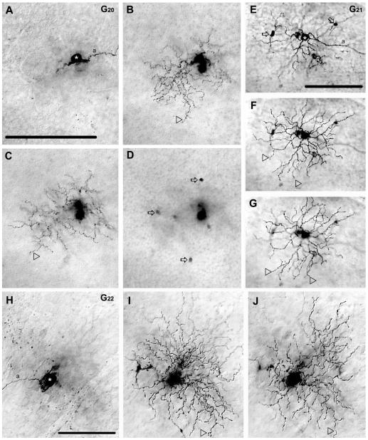
Figure 12.
Morphology and tracer coupling pattern of the G20,G21, and G22 ganglion cell subtypes in the mouse retina. A–D: Photomicrographs show a G20 cell (asterisk) with focal depths on the GCL (A), on sublamina-b of the IPL (B), sublamina-a of the IPL (C), and in the INL (D). Arrowheads point to dendrites that stratify in either sublamina-b (B) or sublamina-a (C) of the IPL. Arrows in panel D point to somata of heterologously coupled amacrine cells in the INL. E–G: Photomicrographs show a G21 cell (asterisk) with focal depths on the GCL (E), on sublamina-b of the IPL (F), and sublamina-a (G) where dendrites stratify (arrowheads). This G21 cell displays tracer coupling to amacrine cells (arrows) with somata displaced into the GCL. H–J: Photomicrographs showing a G22 cell (asterisk) with focal depths on the GCL (H), sublamina-b (I), and sublamina-a (J) of the IPL. The dendritic arbor of this G22 cell appears bistratified with some terminal dendrites (arrowheads) located in sublamina-b (I) or sublamina-a (J). This G22 cell shows no evidence of tracer coupling. a, axon. Scale bars = 100 μm.

