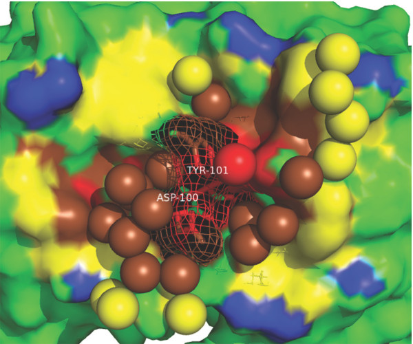
Figure 9.
An example of antibody-antigen interface. The interface between an anti-hen egg white lysozyme antibody D1.3 and a hen egg white lysozyme ([PDB:1VFB], resolution: 1.8 Å, wetness: 0.083, rWBL: 1.143). Only the antibody part (in surfaces) and interfacial water molecules (in spheres) are shown. O0, O1, O2, O3 and non-interface are colored red, brown, yellow, blue and green, respectively. Two residues contribute more than 3.0 kcal/mol [33] are highlighted in mesh and sticks.

