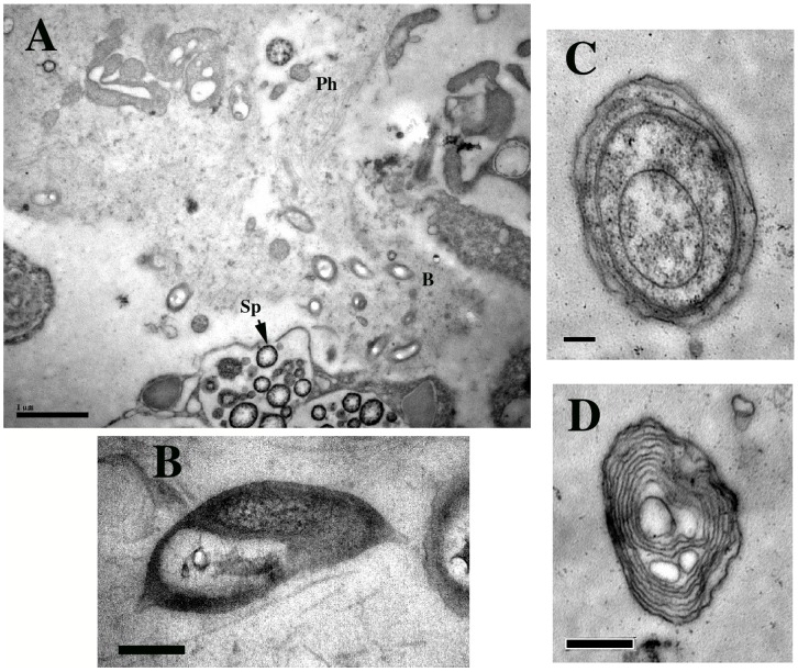
Figure 5. Representative TEM micrographs of Axinella corrugata sponge mesohyl.
A) Wide angle view showing potentially aggregated bacteria (b), possible phage (Ph) and spicule –forming cells (Sp). Scale bar = 1 µm; B) One of several unidentified pear-shaped bacteria within Axinella corrugata sponge mesohyl. Scale bar = 0.2 µm; C) Possible Cyanobacteria, Scale bar = 1 µm; D) Possible Ectothiorhodospiraceae microbial symbiont within Axinella corrugata. Scale bar = 0.5 µm.

