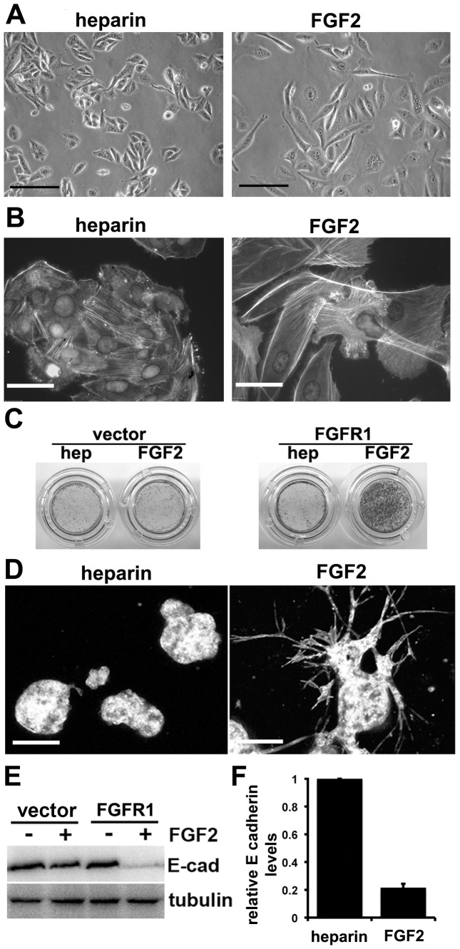
Figure 1. FGFR1 activation promotes EMT in 94-10 cells.
A. 94-10-FR1 cells were cultured with heparin or heparin and FGF2 for 72 h. Images were taken at 72 h (bars = 100 µm). B. At 72 h 94-10-FR1 cells cultured with heparin or heparin and FGF2 were fixed and stained with DAPI and Phalloidin-Alexa 488 (bars = 30 µm). Images represent a merged picture of DAPI and Phalloidin-Alexa 488 staining. C. Transwell assays were used to assess changes in migration in 94-10-FR1 and control cells. Cells which had migrated (cells on the lower part of the transwell) were stained at 36 hr with haematoxylin. D. Invasion assays were performed on 94-10-FR1 cells seeded onto a layer of 50% matrigel containing heparin or heparin and FGF2. At 96 h they were fixed, stained with Phalloidin and imaged using confocal microscopy (bars = 0.5 mm). E. Western blots for E-cadherin and tubulin (loading control) on 94-10-FR1 and 94-10 vector control cells cultured with heparin or heparin and FGF2 for 72 h. F. Real time RT-PCR for E-cadherin on 94-10-FR1 cells cultured with heparin or heparin and FGF2 for 72h. SDHA was used an internal control.

