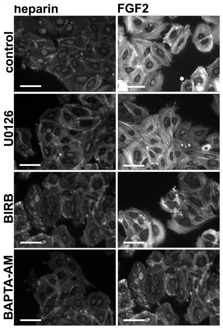
Figure 4. FGFR1-induced cytoskeletal changes are calcium-dependent.
94-10-FR1 cells were cultured with inhibitors for 1 h prior to addition of heparin or heparin and FGF2 for 2 h. Cells were fixed, stained with Phalloidin and DAPI, and imaged (bars = 30 µm). Images represent combined DAPI and Phalloidin staining.

