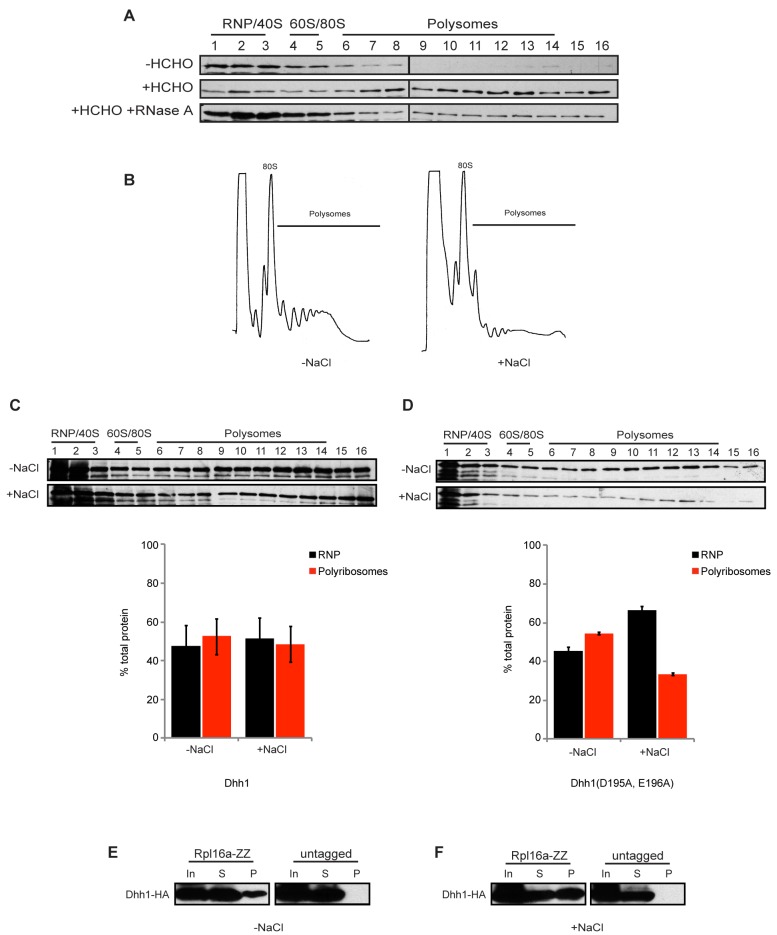
Figure 4. Dhh1 protein associates with slowly translocating polyribosomes.
(A) Extracts from dhh1Δ cells expressing HBHT-tagged Dhh1 were separated by velocity sedimentation on 15%–45% sucrose gradients and protein was extracted from each fraction by TCA precipitation. SDS-PAGE was performed, protein was transferred to PVDF membrane, and Dhh1 was detected by Western blotting with anti-RGS-His antibody. −HCHO, without formaldehyde crosslinking; +HCHO, with formaldehyde crosslinking; +RNase A, with ribonuclease A. (B) Representative polyribosome traces from extracts of cells treated without (−NaCl) and with (+NaCl) 1 M NaCl. (C) Same analysis as in (A) for HBHT-Dhh1 association with polyribosomes from cells treated with or without 1 M NaCl. (D) Same analysis as in (C) of mutant Dhh1(D195A, E196A). (E) Ribosome affinity purification was performed on extracts from crosslinked cells resuspended in media without 1 M NaCl, expressing both RPL16a-ZZ and DHH1-HA or DHH1-HA alone (untagged). Shown is a Western blot probed for Dhh1 using anti-HA antibody. (In, one-tenth input; S, one-tenth supernatant; P, pellet). (F) same analysis as (E), but with cells treated with 1 M NaCl.

