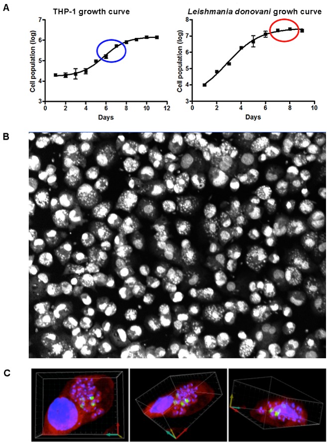
Figure 3. THP-1 infection with L. donovani.

A) Growth curves of THP-1 and L. donovani, with the optimal development points for infection highlighted by blue and red circles, respectively. B) Image acquired with an Opera confocal microscope showing THP-1 infected with L. donovani after Draq5 (DNA) staining. C) 3-D reconstitution of multiple series confocal pictures illustrating from two different perspectives THP-1 macrophages stained with Syto-60 (red) and infected by L. donovani parasites. Dapi was used to stain the DNA (blue) of both the host cells and the parasites. BrdU incorporation detected by immunofluorescence (green) indicates the replication of intracellular amastigote parasites.
