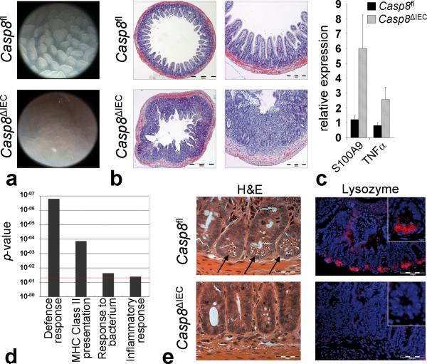
Figure 1. Casp8ΔIEC mice spontaneously develop ileitis and lack Paneth cells.
(a) Representative endoscopic pictures and (b) H&E stained cross sections showing villous erosions in the terminal ileum of Casp8ΔIEC. (c) RT-PCR showing increased level of inflammatory markers in the terminal ileum of Casp8ΔIEC mice (6 mice per group +SEM, relative to HPRT). (d) Gene ontology analysis of genes significantly downregulated in gene chip analysis of IEC from 3 control and 3 Casp8ΔIEC mice. (e) Ileum cross sections stained with H&E and lysozyme for Paneth cells (inset = single crypt at higher magnification). Arrows indicate crypt bottom with Paneth cells.

