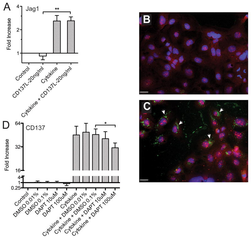
Figure 4. Studies on Caco-2BBe cell gene expression show coordinate regulation of M cell genes Jagged1 and CD137.
A. TNFα plus LTβR agonist (“Cytokine”: 100ng/ml TNFα and 5μg/ml of LTβR agonist antibody) induced upregulation of Jagged1 with no additional influence of soluble CD137L agonist. (t-test analysis; **, P<0.005)
B. Confocal images of untreated Caco-2BBe cells stained for CD137 (green) and Jagged1 (red). Nuclei are blue. (Scale bar: 30 μm)
C. Confocal images of cytokine treated Caco-2BBe cells shows upregulation of CD137 (arrows), but there is no co-localization of CD137 (green) with Jagged1 (red). (Scale bar: 30 μm)
D. Cytokine treatment of Caco-2BBe cells induces expression of CD137, but expression shows a dose-dependent inhibition by the Notch signaling inhibitor DAPT. One tailed paired t- test comparing DMSO control to 100μm DAPT was significant (P<0.05).

