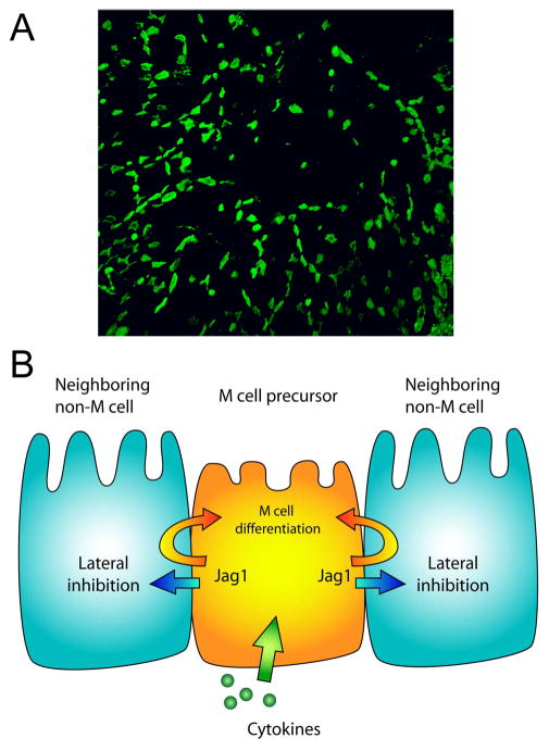
Figure 5. Model of Jagged-Notch interactions and M cell patterns in Peyer’s patch epithelium.
A. A low magnification view of the overall distribution of M cells (green) across Peyer’s patch follicle epithelium. Note that at this low magnification, dispersion of M cells derived from crypt stem cell progenitors is evident, though the rare M cell clusters are not as visible.
B. Simplified cartoon model of M cell editing by Jagged1 and Notch interactions, indicating a possible role for Jagged1 in both lateral inhibition of M cell development and cis-reinforcement of M cell lineage decisions.

