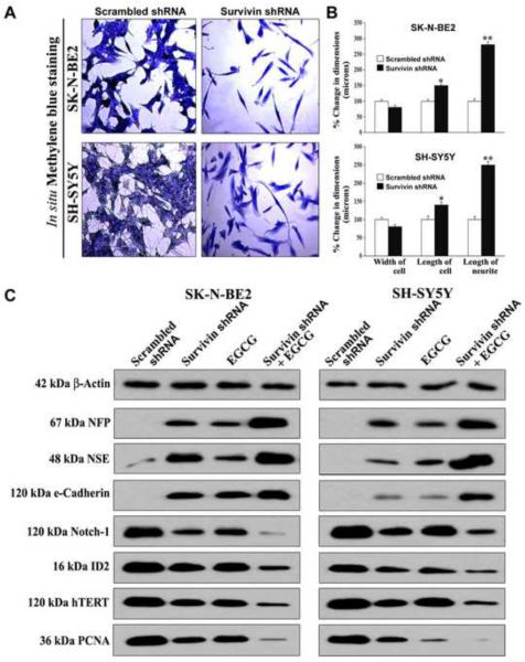
Fig. 3.
Down regulation of survivin inhibited cell proliferation and induced morphological and biochemical features of neuronal differentiation. (A) In situ methylene blue staining showed inhibition of cell growth and increase in morphological features of neuronal differentiation. Photographs were taken after transfection (72 h) with plasmid vector carrying survivin shRNA cDNA (0.5 μg/ml) or scrambled shRNA cDNA (0.5 μg/ml). (B) Measurement of morphological features of neuronal differentiation (width of cell, length of cell, and length of neurite extension). Mean values of 3 independent experiments are shown (*p < 0.05; **p < 0.01). (C) Representative (n = 3) Western blots to show changes in biochemical markers of neuronal differentiation and cell proliferation. Treatments: transfection with plasmid vector carrying scrambled shRNA cDNA (0.5 μg/ml) for 48 h, transfection with plasmid vector carrying survivin shRNA cDNA (0.5 μg/ml) for 48 h, 50 μM EGCG for 24 h, and survivin shRNA (0.5 μg/ml) for 48 h + 50 μM EGCG for last 24 h. Expression of β-actin was used as a loading control.

