
Figure 3. Expression and localization of BCRP in human brain tumor biopsy specimens.
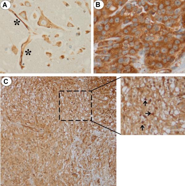
(A) Immunohistochemical analysis of BCRP in healthy, control human brain biopsy. BCRP staining of brain endothelial cells (denoted as *) and significant cytoplasmic/membraneous expression without nuclear expression of BCRP in brain glial cells; (B) A strong BCRP signal was observed in the cytoplasmic/membraneous compartment of a human breast cancer specimen. No nuclear BCRP staining was noted; (C) Biopsy GBM specimen at low (200X) and high (400X) magnification (inset) demonstrating nuclear BCRP staining in a subpopulation of tumor cells (denoted by arrows).