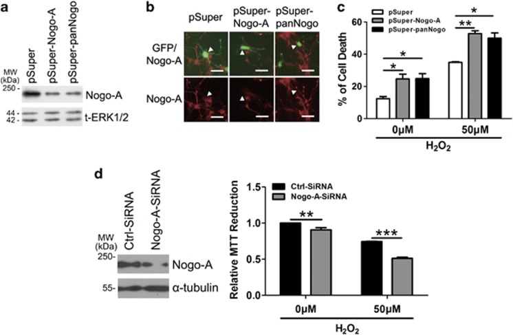
Figure 3.
Downregulation of Nogo-A increases the neuronal susceptibility to oxidative stress. (a) Rat-derived PC12 cells transfected with pSuper, pSuper-Nogo-A and pSuper-panNogo for 48 h were lysed and immunoblotted with polyclonal antibody (pAb) A620. Total extracellular signal-regulated kinase 1/2 (t-ERK1/2) was selected as a loading control. (b) Cortical neurons co-transfected (arrow head) with pEGFP-N1 and the small hairpin RNA (shRNAs) above for 48 h were immunostained with pAb A620. (c) After transfection for 48 h as described in (b), neurons were treated with 0 or 50 μM hydrogen peroxide (H2O2) for 12 h, cell death rate was determined by propidium iodide (PI) (+)/EGFP (+). (d) Neurons were transfected with ctrl-small interfering RNA (siRNA) and Nogo-A-siRNA for 48 h, and cell lysates were blotted with pAb A620 (left). After transfection for 48 h as described in (d), 0 or 50 μM H2O2 was added into cell culture for another 12 h, cell death rate was determined by 3-(4,5-dimethyldiazol-2-yl)-2,5-diphenyltetrazolium bromide (MTT) assay. Bar=50 μm. n=3, mean±S.D., one-way analysis of variance (ANOVA), *P<0.05; **P<0.01; ***P<0.001

