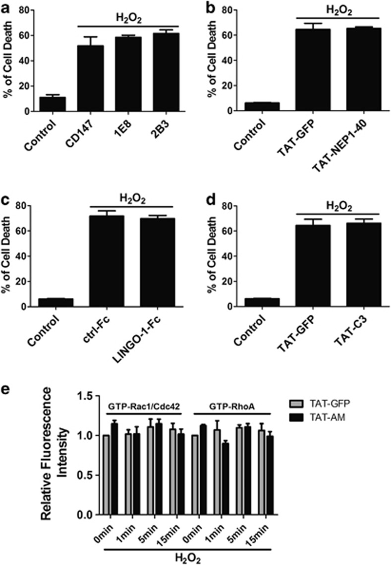
Figure 4.
Nogo-66-NgR/LINGO-1 and RhoA-ROCK signaling pathways are not involved in the neuroprotection. 1E8 and 2B3 (a), HIV-1 trans-activating (TAT)-NEP1–40, Nogo extracellular peptide, residues 1–40 (b), LINGO-1-Fc (c) and RhoA inhibitor TAT-C3 transferase (TAT-C3) (d) were added to cell culture, followed by 50 μM hydrogen peroxide (H2O2) incubation for 12 h in the presence of these proteins, cell death assay was performed by propidium iodide (PI) (+)/Hoechst (+). CD147 antibody, TAT-GFP and ctrl-Fc protein were selected as controls, respectively. (e) Active Rac1/Cdc42 and RhoA were detected by glutathione S-transferase (GST)-PBD and GST-RBD using in situ GST pull-down assay for neurons incubated in 50 μM H2O2 for indicated time with TAT-AM or not and then quantified. n=4, mean±S.D.

