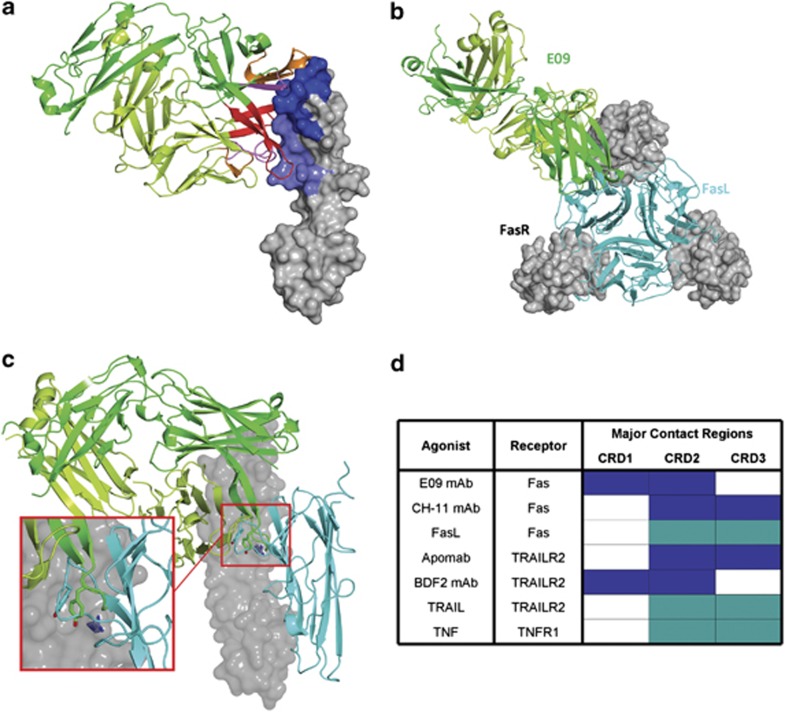
Figure 2.
Structural analysis of Fas agonist antibody E09. (a) The structure of Fas ECD in complex with E09 Fab. Fas ECD is represented with a surface rendering and the epitope on CRD1 and CRD2 coloured in darker blue and lighter blue, respectively. The E09 is shown as a cartoon with the heavy chain in darker green and the light chain in lighter green. The CRD1, 2 and 3 are coloured in light pink, dark pink and red, respectively. (b) Model of a trimeric Fas-FasL complex that shows the binding of E09 in green and FasL in cyan in top view. (c) A representation of Fas ECD in complex with E09 Fab in which the epitope overlap between FasL and E09 is visible. A single tyrosine in FasL (Y218) or E09 (VH_100d) makes contact with Fas_R86 (zoom). (d) Comparative analysis of broad epitopes on death receptors covered by natural ligands and agonistic antibodies

