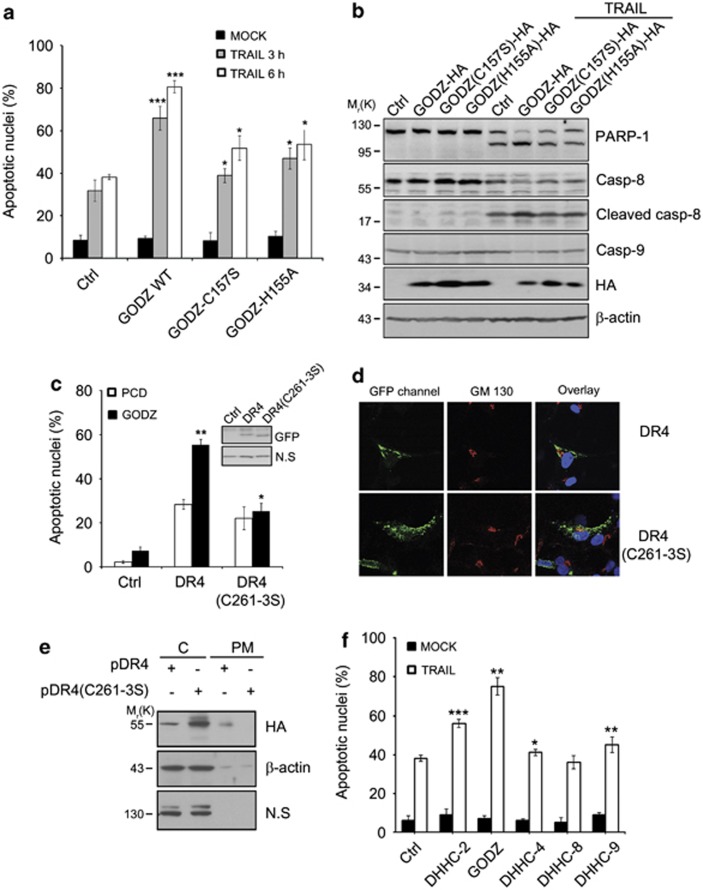
Figure 5.
Contribution of GODZ DHHC motif to plasma membrane (PM) targeting of DR4 and TRAIL sensitivity. (a) Mutation in GODZ DHHC motif loses the ability to stimulate TRAIL-mediated cell death. HeLa cells were transiently co-transfected with pEGFP (Clontech) and either pcDNA3-HA (Ctrl), pGODZ WT-HA, pGODZ(C157S)-HA, or pGODZ(H155A)-HA for 24 h, and then left untreated (MOCK) or treated with TRAIL (100 ng/ml) for the indicated times. After staining with EtHD, cell death was measured by counting the number of both GFP and EtHD-positive cells among total GFP-positive cells. Bars indicate mean±S.D. (n=3). P-values were estimated using t-test versus control (*P<0.05; **P<0.01; ***P<0.001). (b) Lack of caspase-8 activation by GODZ mutants. HeLa cells were transiently transfected with pcDNA3-HA, pGODZ-HA, pGODZ(C157S)-HA, or pGODZ(H155A)-HA for 24 h, and left untreated or treated with TRAIL (100 ng/ml) for 3 h. Cell extracts were prepared and analyzed by western blotting using the indicated antibodies. (c) Mutation in cysteine-rich motif of DR4 (DR4C261-3S) weakens the pro-apoptotic activity. HeLa cells were transfected with pcDNA3 (PCD; an empty vector) or pGODZ, and pEGFP (Clontech) (Ctrl), pDR4-GFP, or pDR4 (C261-3S)-GFP for 18 h, and cell death ratios were measured as described in (a). Bars indicate mean±S.D. (n=3). P values were estimated using t-test versus control (*P<0.05; **P<0.01). (d) Failure in the targeting of DR4 C261-3S mutant to PM. COS7 cells were transiently transfected with pDR4-GFP or pDR4 (C261-3S)-GFP for 12 h. Thereafter, cells were fixed and stained with anti-GM130 antibody (red). Green and red fluorescent images were acquired under a confocal microscope (Zeiss, Thronwood, NY, USA) and then overlaid with Hoechst 33258 staining. (e) Fractionation assay showing the impaired targeting of DR4 C261-3S mutant to PM. HEK 293T cells were transiently transfected with pDR4-HA or pDR4 (C261-3S)-HA for 24 h, and then cell extracts were subjected to fractionation assay to separate the PM from the cytosol (C), as described in materials and methods. The fractions were analyzed by western blotting with anti-HA and anti-β-actin antibodies. (f) Effects of various DHHC motif-containing proteins on TRAIL sensitivity. HeLa cells were transiently transfected with pEGFP (Clontech) and various DHHC motif-containing constructs for 24 h, and then left untreated (MOCK) or exposed to TRAIL (100 ng/ml) for 3 h. Cell death ratios were measured as described in (a). Bars indicate mean±S.D. (n=3). P-values were estimated using t-test versus control (*P<0.05; **P<0.01; ***P<0.001). The color reproduction of this figure is available at the Cell Death and Differentiation journal online

