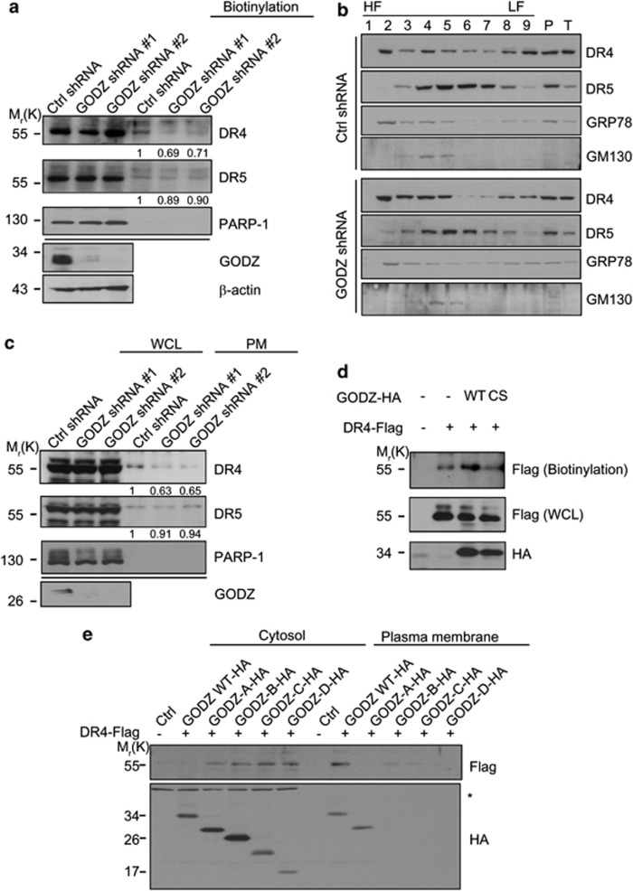
Figure 6.
Depletion of GODZ expression reduces PM-localized DR4. (a) Biotinylation assay showing GODZ-dependent localization of DR4 to the PM. HeLa/Ctrl shRNA and HeLa/GODZ shRNA stable cells were exposed to biotin, and the PM fractions were prepared by fractionation and subjected to biotinylation assays with Sulfo-NHS-SS-Biotin (Pierce biotechnology). Western blotting was then performed with anti-GODZ, anti-DR4, anti-DR5, and anti-β-actin antibodies. Densitometric analysis of western blots was performed using a multi-gauge imaging system and their relative ratios to that of control shRNA are represented. (b and c) Fractionation assay using gradient centrifugation shows impaired membrane targeting of DR4 by GODZ deficiency. HeLa/Ctrl shRNA and HeLa/GODZ shRNA cell extracts were subjected to discontinuous gradient centrifugation using OptiPrep (Sigma) (iodixanol) as a gradient medium. After centrifugation, nine fractions of equal volume (500 μl each) were concentrated and subjected to western blotting using anti-DR4, anti-DR5, anti-GRP78, and anti-GM130 antibodies (b). The fractions seven, eight, and nine were pooled and subjected to western blotting using anti-DR4, anti-DR5, and anti-PARP-1 antibodies. Densitometry analysis of western blots was performed and relative ratios of DR4 and DR5 signals to control are represented (c). (d) Enhanced targeting of DR4 to cell surface by GODZ but not by GODZ C157S mutant. HEK 293T cells were transfected with pDR4-Flag and either pGODZ-HA or pGODZ(C157S)-HA for 24 h, and then subjected to biotinylation assay with (Pierce biotechnology) as described in (a). Western blotting was performed with anti-Flag and anti-HA antibodies. (e) Effects of GODZ mutants on targeting of DR4 to PM. HEK 293T cells were co-transfected with pDR4-Flag and either pGODZ-HA or GODZ deletion mutant for 24 h. The PM and cytosol fractions were prepared as described in materials and methods, and subjected to western blotting using anti-Flag or anti-HA antibody. The asterisk in (e) indicates nonspecific signal of the cytosol fraction. HF, high-density fraction; LF, low-density fraction; P, pallet; T, total

