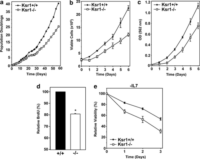
Figure 2.
Pre-B cells lacking Ksr1 exhibit reduced proliferation and increased apoptosis. (a) Bone marrow from Ksr1+/+, Ksr1+/−, and Ksr1−/− littermates was placed into media containing IL-7 on day 0. Viable cells were counted at intervals by Trypan Blue Dye exclusion assay. Population doublings were calculated. A representative experiment is shown; similar results were obtained with bone marrow from littermates from different parents. (b and c) Equal numbers of Ksr1+/+ and Ksr1−/− pre-B cells were plated in triplicate and the total number of viable cells was assessed by Trypan Blue Dye exclusion (b) or MTT assay (c) for 6 days. (d) Ksr1+/+ and Ksr1−/− pre-B cells were pulsed with BrdU, and incorporation of BrdU was determined by flow cytometry. Data are expressed as average BrdU incorporation relative to Ksr1+/+ cells (n=4 of each); P<0.0001. (e) Equal numbers of Ksr1+/+ and Ksr1−/− pre-B cells were cultured in media lacking IL-7 beginning at day 0. Viability was monitored for 3 days by Trypan Blue Dye exclusion assay. Percent viability at each time is expressed relative to the viability at time 0

