
Fig. 5.
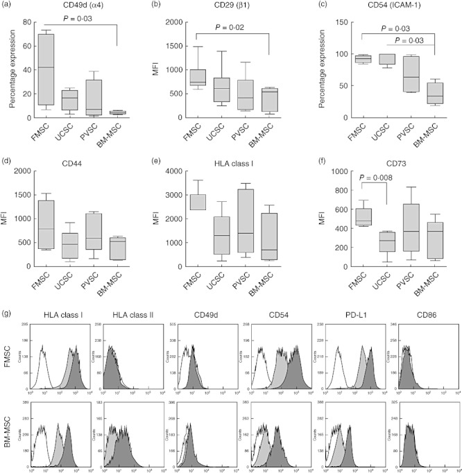
Fetal membrane stromal cells express high levels of adhesion molecules. (a–f) Expression of different markers on stromal cells from fetal membrane (FMSCs), umbilical cord (UCSCs), placental villi (PVSCs) and bone marrow-derived mesenchymal stromal cells (BM-MSCs) were analysed by flow cytometry (n = 4 for each type of stromal cell). The Mann–Whitney U-test was used to compare the mean fluorescence intensity (MFI) or the percentage expression of cell surface markers on different types of stromal cells. The data are presented as box-and-whiskers with the maximum, minimum, median, 25th and 75th quartiles. (g) Representative histograms of expression of different markers on unstimulated (light grey histograms) or interferon (IFN)-γ-stimulated (dark grey histograms) FMSCs (n = 6) and BM-MSCs (n = 3). White histograms depict isotype controls.