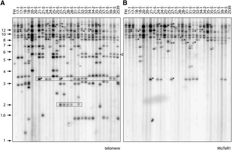
Figure 6 .
Segregation of telomere instability in a genetic cross. DNA was extracted from single-spore cultures of each progeny isolate (G1). These isolates were subcultured for four additional generations, new single-spore cultures (G5) were established, and DNA was extracted from the resulting cultures. The genomic DNA samples were digested with PstI, fractionated by electrophoresis, and electroblotted to a membrane. The membrane was probed sequentially with 32P-labeled telomere and MoTeR1 fragments. The left and right lanes contain the DNAs from the parental strains. DNA samples from the G1 and G5 subcultures of an individual progeny isolate were loaded in adjacent lanes to allow comparison of their telomeric restriction fragment profiles. Molecular sizes are shown on the left. The white arrowhead marks 2539TEL10, a MoTeR2-containing telomere that was highly stable. (A) Phosphorimage of the TTAGGG-probed gel. Boxes outline fragments that segregated among the progeny but that were not present in either parent. (B) The same membrane used in A was stripped and reprobed with a fragment from the MoTeR1 RT. Asterisks mark hybridizing fragments that were present in the G0 culture but absent in the G5. Open circles show fragments that appeared in the G5 cultures.

