Figure 3.
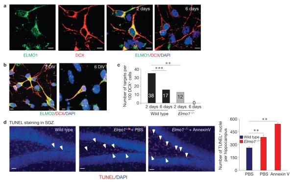
ELMO1-dependent phagocytosis of DCX+ cells in vitro. (a) Representative images of primary hippocampal neuronal cultures grown for 2 or 6 days in vitro immunolabelled for DCX and ELMO1 (for n > 30 fields). Scale bars, 5 μm. (b) Primary hippocampal neuronal cultures grown for 2 or 6 days in vitro (DIV) were immunolabelled for DCX and ELMO2. Representative confocal microscopy images (for n > 30 fields) are shown. The expression level of ELMO2 was not altered under these conditions in the DCX+ cells after 2 or 6 days in culture. Scale bars, 5 μm. (c) Cultures of neurons from a were fed with targets (carboxylate-modified beads) and assessed for engulfment. The bars represent quantifications of phagocytosed targets by primary DCX+ cells from wild-type and Elmo1−/− mice after 2 or 6 days in culture. A one-way analysis of variance with a Newman–Keuls multiple comparison test was carried out for n = 100 DCX+ cells per group (*** P < 0.01; *** P < 0.001). (d) Accumulation of apoptotic nuclei in PBS- or long-term annexin V-treated Elmo1−/− mice, compared with wild-type littermates, was examined by TUNEL immunolabelling (indicated by white arrowheads) of the SGZ. Representative images from wild-type and Elmo1−/− mice are shown. The bar graph represents a quantification of TUNEL-positive nuclei (mean ± s.e.m.). Student’s t -test was carried out for n 5 mice per group, with at least ten slices analysed for each mouse (** = P < 0.01). Scale bars, 50 μm.

