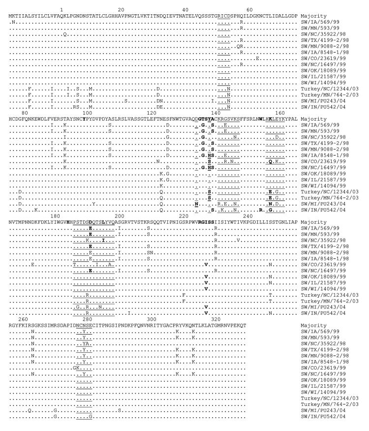
Figure 2.
Alignment of deduced amino acid sequences within the hemagglutinin (HA) 1 region of HA genes of H3N2 swine influenza viruses (SIVs), H3N2 turkey isolates, and H3N1 SIVs. The amino acid sequence represents the consensus sequence, and the amino acid at position 1 is the first amino acid following the signal peptide (37). Dots represent amino acids similar to the consensus. Note that according to H3 structure (37), the residues representing the antigenic sites are underlined and the receptor binding sites are in boldface. The alignment shows that PU243 and PU542 may have emerged from the H3N2 turkey isolates. The residues within the receptor-binding site are relatively conserved.

