FIGURE 1.
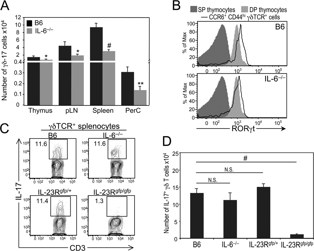
Phenotypic and functional analysis of γδ-17 cells in interleukin (IL)-6–deficient mice. (A) Comparison of the numbers of γδ-17 cells, defined as CCR6+ and CD44hi, in the thymus, peripheral lymph nodes (pLN), spleen, and peritoneal cavity (PerC) of 5- to 6-week-old C57BL/6 (B6) and IL-6−/− mice. *P ≤ 0.05; **P ≤ 0.01; #P ≤ 0.001. (B) Histograms showing representative staining of RORγt in wild-type and IL-6–deficient γδ-17 cells from pLN. RORγt expression levels on CD4+ and CD8+ single-positive (SP) thymocytes are shown as a negative control, whereas those on CD4+CD8+ double-positive (DP) thymocytes are shown as a positive control. (C) Six-week-old B6, IL-6−/−, IL-23Rgfp/+ (IL-23R+/−), and IL-23Rgfp/gfp (IL-23R−/−) were infected intraperitoneally with a 0.3 LD50 dose of wild-type Listeria (3 × 10⁵ pfu). Five days after infection, splenocytes were harvested and restimulated in vitro with IL-1, IL-23, and Pam3Cys (a TLR2/1 agonist) for 4 hours in the presence of brefeldin A. Representative dot plots show IL-17 versus CD3 staining in gated γδ T cells. Numbers represent percentage of IL-17+ γδ T cells. (D) Summary of data presented in C. # P ≤ 0.001. N.S., not significant.

