Figure 5. .
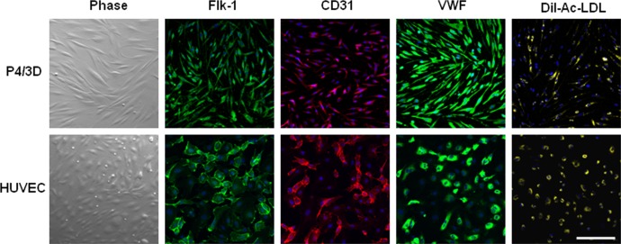
Differentiation into vascular endothelial cells. Single cells of P4/3D aggregates were cultured on plastic in EGM2 with VEGF for 3 days, yielding spindle cells similar to HUVEC. They uniformly expressed Flk-1, CD31, and von Willebrand factor, and took up Dil-Ac-LDL (top) in a similar fashion to the positive control of HUVEC cultured in the same condition (bottom). Nuclei were counterstained by Hoechst 33342 (blue). Scale bar = 100 μm.

