Figure 6. .
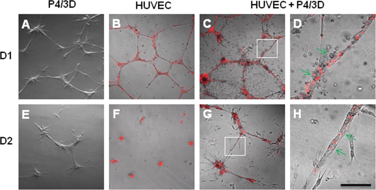
Support of HUVEC vascular tube network. Fluorescence pre-labeled (red) HUVEC and P4/3D cells were seeded alone or together on the surface of 100% Matrigel in EGM2. Although vascular tube-like network was noted in all three conditions at Day 1 (A–C), such network in P4/3D cells (A) or HUVEC (B) alone was disintegrated by Day 2 (E, F). In contrast, the network formed by cocultured P4/3D cells and HUVEC (C) was maintained at Day 2 (G) and Day 5 (not shown). High magnification of insets (C, G) revealed close association between P4/3D cells and HUVEC (red) in the network at Day 1 (D) and Day 2 (H). Scale bar = 200 μm for A–C and E–G, and 50 μm for D and H.

