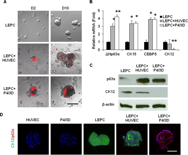
Figure 7. .
Epithelial sphere growth in 3D Matrigel. LEPCs derived from dispase-isolated epithelial sheets alone or mixed with fluorescence prelabeled (red) HUVEC or P4/3D cells to generate sphere growth from Day 2 to Day 10 in 3D Matrigel (A). Compared with those formed by LEPC alone, spheres formed by LEPC+HUVEC and by LEPC+P4/3D expressed significantly more ΔNp63α, CK15, and CEBPδ transcripts (B, n = 3, *P < 0.05, **P < 0.01). In addition, expression of CK12 transcripts by LEPC+HUVEC was not different from that of LEPC alone (B, n = 3, P > 0.05), whereas that by LEPC+P4/3D was not detectable (B, n = 3, P < 0.01). Compared with LEPC alone, expression of p63α protein was elevated in both LEPC+HUVEC and LEPC+P4/3D (C, n = 3, P < 0.05). In contrast, expression of CK12 protein was not reduced in LEPC+HUVEC (C, n = 3, P > 0.05) but was reduced to a nondetectable level in LEPC+P4/3D using β-actin as a loading control (C, n = 3, P < 0.05). Double staining with p63α and CK12 showed that HUVEC or P4/3D cells alone did not expresss p63α or CK12, whereas CK12 was expressed by LEPC alone and LEPC+HUVEC but not LEPC+P4/3D (D). Scale bar = 200 μm for A and 100 μm for D.

