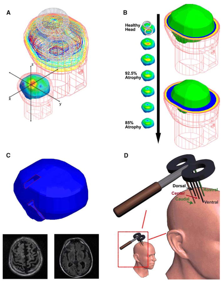
Fig. 1. Geometries.

a Healthy head model and the model coordinate system: This image depicts the Wnite element mesh borders of the skin (flesh color), skull (yellow), CSF (light blue), gray matter (dark blue), and white matter (red). The coordinate system is shown in the foreground image and was used for all the models. b Increasing symmetric atrophy models: the models are displayed from the healthy head model to the 85% atrophy model. On the right side, the healthy head model (upper) and the 85% atrophy model (lower) are shown to highlight the increasing thickness of the CSF and the decreasing cortical thickness. The skin mesh is shown in the flesh color where the tissue thicknesses are highlighted in the transverse slices; scalp (flesh colored), skull (yellow), CSF (blue), brain (bright green). Notice the increasing CSF thickness and the decreased cortical size between the two models. c. Widened sulci model: the base sulci model is shown with the widened sulcal regions highlighted and sample MRI slices. d Evaluation line locations: the lines were located with the center line normal to the figure-of-eight coil center and the other lines 1 cm ventral, dorsal, anterior, and posterior to the center line (note that the figure is not drawn to scale but with the lines and the coil drawn to highlight their placement)
