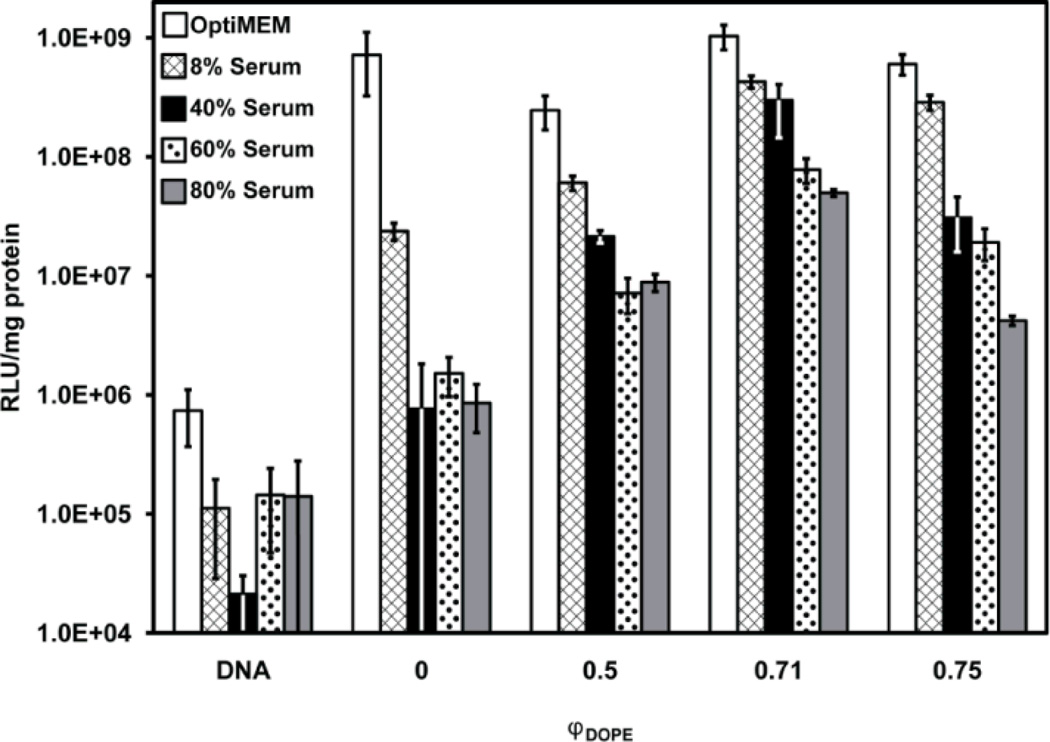
Fig. 3.
Influence of serum on normalized luciferase expression in COS-7 cells treated with lipoplexes formed from pCMV-Luc and mixtures of reduced BFDMA and DOPE for 4 h. The final concentration of BS is given in the legend. DNA was present at a concentration of 2.4 µg/ml for all samples. “DNA” denotes a control with DNA only (no lipid). Molar fractions of DOPE, φDOPE = DOPE/(BFDMA+DOPE), are given on the x-axis for each sample. The concentration of BFDMA in each sample was 8 µM. Luciferase expression was measured 48 h after exposure to lipoplexes. Error bars represent one standard deviation.

