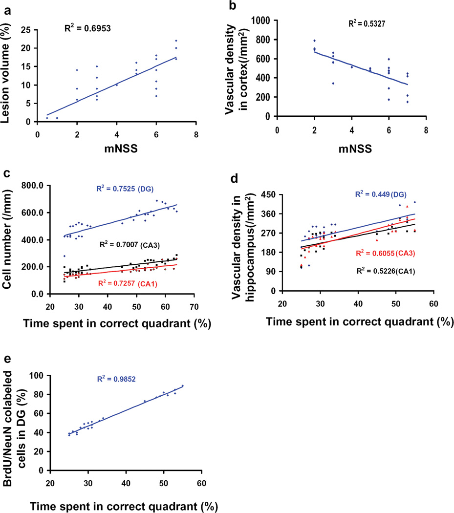
Fig. 7.
Correlation of functional outcomes with lesion volume, cell loss, angiogenesis, and neurogenesis. Data from all four groups were included to generate the correlations between functional and histological outcomes. The top panel line graphs show that the functional outcomes (mNSS scores) are significantly and positively correlated with the lesion volume (a; P<0.05) but inversely correlated with the vascular density (b; P<0.05). The other panel line graphs show that spatial learning performance is significantly and positively correlated with the number of neuron cells (c), vascular density (d), and NeuN/BrdU-positive cells (e) in the ipsilateral hippocampus measured at day 35 in rats after TBI and EPO treatment (P<0.05). Data represent mean ± SD. n (rats/group) = 8.

