FIGURE 5:
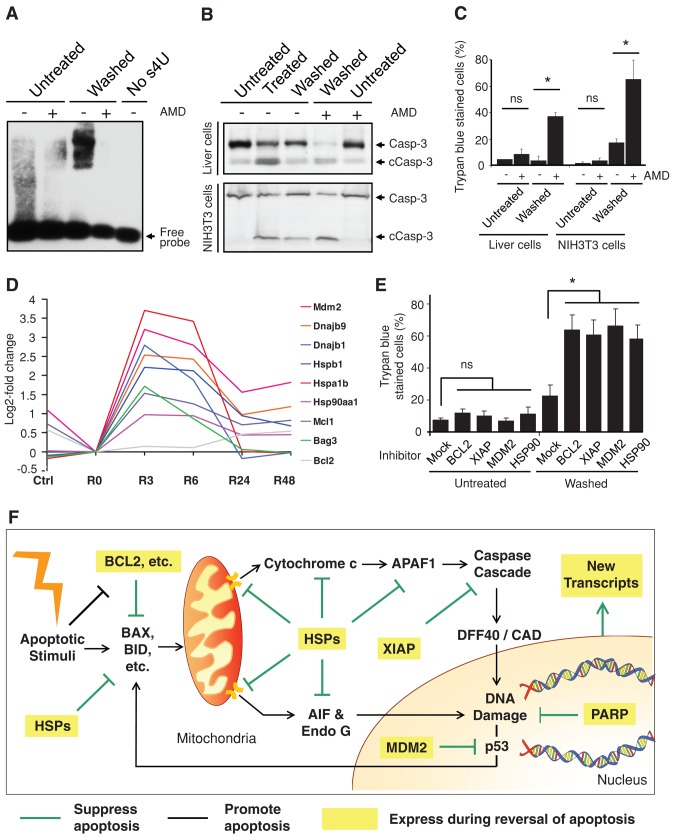
Critical contributing factors in reversal of apoptosis. (A) RNA blot for detecting new RNA synthesis in untreated vs. treated and washed (Washed) liver cells 1 h after a 5-h exposure to 4.5% ethanol and with and without transient exposure (1 h, 1μg/ml) to actinomycin D (AMD). No s4U served as negative control for the probe binding to RNA. The detailed procedure is described in Materials and Methods. (B) Western blot analysis of the cleavage of caspase-3 (Casp-3) on the total lysate of untreated, treated (5-h exposure to 4.5% ethanol for liver cells, 20-h exposure to 10% DMSO for NIH 3T3 cells), and washed liver cells and NIH 3T3 cells with and without transient exposure to actinomycin D (AMD) immediately after removal of apoptotic stimuli. The AMD-exposed cells were cultured in fresh medium for 23 h and then subjected to the analysis. c, cleaved form. (C) Percentage of untreated as well as treated and washed liver cells and NIH 3T3 cells with and without transient AMD exposure that displayed plasma membrane permeability in trypan blue exclusion assay. The AMD exposed-cells were cultured in fresh medium for 23 h and then subjected to the assay. ns, p > 0.05; *p < 0.05; n = 3 independent experiments. Error bars denote SD. (D) A time-course microarray study in liver cells, log2-fold change of gene expression comparison between ethanol-induced apoptotic cells (R0), untreated cells (Ctrl), and induced cells that were then washed and further cultured in fresh medium for 3 (R3), 6 (R6), 24 (R24), and 48 h (R48). The log2 signal values from three biological replicates were averaged (geometric mean) for each time point. (E) Percentage of untreated and washed liver cells with and without 24-h exposure of inhibitors of BCL-2 (ABT 263, 1 μM), XIAP (Embelin, 20 μM), MDM2 (Nutlin-3, 10 μM), and HSP90 (17-allylaminogeldanamycin, 0.5 μM) that displayed full plasma membrane permeability in trypan blue exclusion assay. The corresponding cells were exposed to the inhibitors after apoptotic stimuli had been removed from the treated cells and then further cultured for 24 h. ns, p > 0.05; *P < 0.05; n = 3 independent experiments. Error bars denote SD. (F) Proposed model for reversal of apoptosis. Expression of multiple prosurvival factors and new transcripts during reversal of apoptosis promotes cell survival by suppressing the activated apoptotic pathways and repairing the cells.

