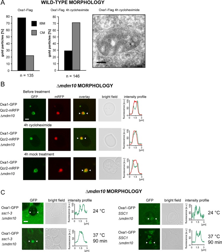
FIGURE 4:
The predominant localization of Oxa1 in the IBM under fermentable growth conditions depends on the import of nuclear-encoded proteins. (A) Quantitative immuno-EM of the distribution of Oxa1-Flag in cells treated for 4 h with (middle) or without (left) cycloheximide, an inhibitor of cytoplasmic translation. The cells were grown in galactose-containing medium. Right, representative image. The arrowhead points to a gold particle. (B) Live-cell fluorescence microscopy of Δmdm10 cells expressing Oxa1-GFP and Qcr2-mRFP. The cells were grown on glucose-containing agar plates and transferred into fresh liquid growth medium containing glucose. They were imaged before (top) and after 4 h treatment with cycloheximide. As a control, the cells were incubated for 4 h with the solvent DMSO. The intensity profiles show the distribution of the fluorescence signals between the arrowheads. (C) Distribution of Oxa1-GFP in ssc1-3 cells exhibiting enlarged mitochondria due to a lack of MDM10. In ssc1-3 cells, mitochondrial import is largely normal at the permissive temperature (24°C), whereas at the nonpermissive temperature (37°C) import is inhibited. Left, distribution of Oxa1-GFP in ssc1-3 cells grown in glucose-containing medium at both temperatures. Right, control experiments with SSC1 cells. Scale bars, 100 nm (A) and 2 μm (B, C).

