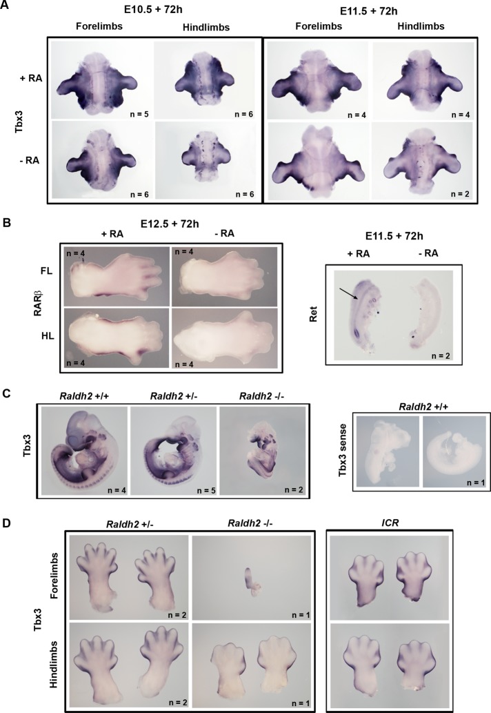
FIGURE 4:
RA activates Tbx3 expression during mouse embryonic development. (A) Whole-mount in situ hybridization with Tbx3 probe of forelimb and hindlimb explants from E10.5 and E11.5 ICR mouse embryos cultured in serum-free medium with serum extender, with or without 200 nM all-trans and 9-cis RA for 72 h. (B) Left, whole-mount in situ hybridization with RARβ probe of handplates and footplates from E12.5 embryo forelimbs (FL) and hindlimbs (HL) cultured with or without RA. Right, whole-mount in situ hybridization with c-Ret probe of genital tracts from E11.5 embryos cultured with or without RA. Arrow indicates the Wolffian duct. (C) Left, whole-mount in situ hybridization of Raldh2 wild-type and mutant E10.5 embryos using antisense Tbx3 probe. Right, whole-mount in situ hybridisation of a wild-type E10.5 embryo with a sense Tbx3 probe. Note that this embryo was damaged in dissection, and therefore two halves are shown. Genotypes are as indicated. (D) Whole-mount in situ hybridization showing Tbx3 expression in handplates and footplates from E13.5 Raldh2+/− and Raldh−/− embryos. Right, handplates and footplates from a stage-matched E13.5 ICR embryo for comparison.

