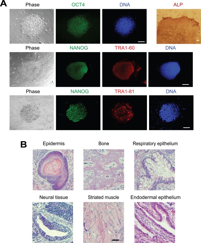
Figure 5. Characterization of LF-iPSCs with immunofluorescence staining and a teratoma formation assay.
(A) Immunofluorescence staining and alkaline phosphatase staining of LF-iPSCs on day 140. Bar, 100 µm. (B) Haematoxylin and eosin staining of histological sections of teratomas derived from LF-iPSCs. Bar, 200 µm.

