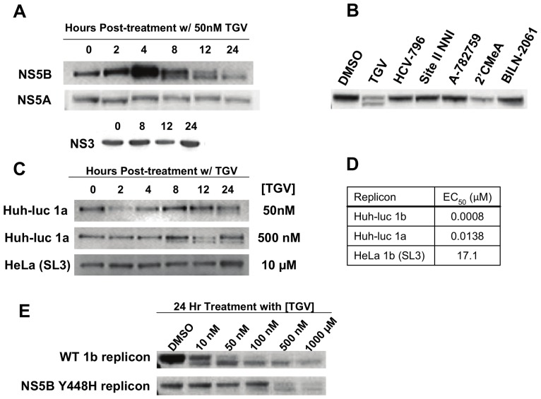
Figure 1. TGV treatment leads to formation of an NS5B double band detectable by Western analysis.
A) NS5B, NS5A, and NS3 Western blots of lysates from 1b (Con-1) replicon cells treated with 50 nM TGV for various periods of time. B) NS5B Western blot of lysates from 1b replicon cells treated for 24 hours with known HCV inhibitors at 50× EC50 concentrations. C) NS5B Westerns of cell lysates collected at various times from Huh-luc 1a replicon cells treated with 50 nM or 500 nM TGV and lysates from HeLa 1b replicon cells treated with 10 µM TGV. D) TGV EC50 values in Huh-luc 1a, 1b, and HeLa 1b (clone SL3) replicon cell lines. E) Western blot of lysates from Lunet cells transiently transfected with wild-type or NS5B Y448H mutant 1b Pi-Rluc replicons and treated with varying concentrations of TGV for 24 hours.

