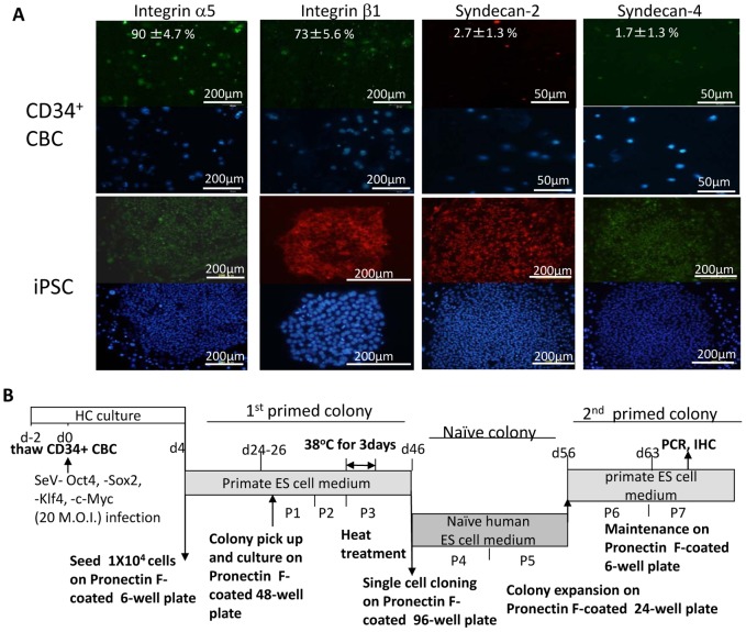
Figure 1. Expression of surface molecules on CD34+ cells and iPSCs.
(A) Adhesion molecules integrin α5, β1, syndecan-2, and -4 on CD34+ CBCs (upper panels) and iPSC colonies (lower panels) detected by immunostaining with the relevant antibody. Alexa 594- and Alexa 488-conjugated secondary antibodies (red and green, respectively) were used to visualize the staining. Nuclei were stained with DAPI (lower photos). Means of the percentages of positive cells with standard deviation are appended in the right top of the photos. (B) Protocol for generation of iPSCs from CD34+ CBCs on Pronectin F-coated dishes with temperature sensitive SeV vectors. P: passage.

