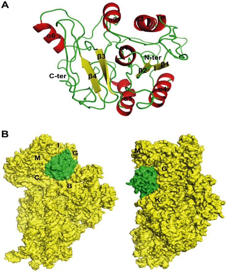
Figure 1. Predicted 3D-structure of DATIN and its docking simulation with 30S ribosomal subunit.
A. Predicted 3D-structure of DATIN. The protein secondary structure elements were labelled and colored (helices and sheets displayed in red and yellow, respectively). B. Docking study of DATIN with 30S ribosomal proteins using PatchDock and FireDock. The protein chains of 30S ribosome and DATIN presented as electrostatic surface and colored in yellow and green, respectively.

