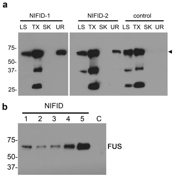
Fig. 2.
FUS in NIFID is relatively insoluble. Gray matter from the frontal cortex of five NIFID cases (lanes 1 to 5) and a control patient (C) was sequentially extracted with buffers of increasing extraction strength. Each extract, corresponding to 20 mg of initial wet tissue weight, was resolved by SDS-PAGE and immunoblotted with antibodies to FUS. There is a consistent increase in insolubility of FUS in NIFID: normal FUS was present in the soluble fractions of NIFID and control (arrowhead), but insoluble FUS (urea fraction) was only present in NIFID cases and not the control (a). Urea-soluble FUS was present in all NIFID.1-5 cases but not in the control brain tissue (NL) (b).

