Figure 4.
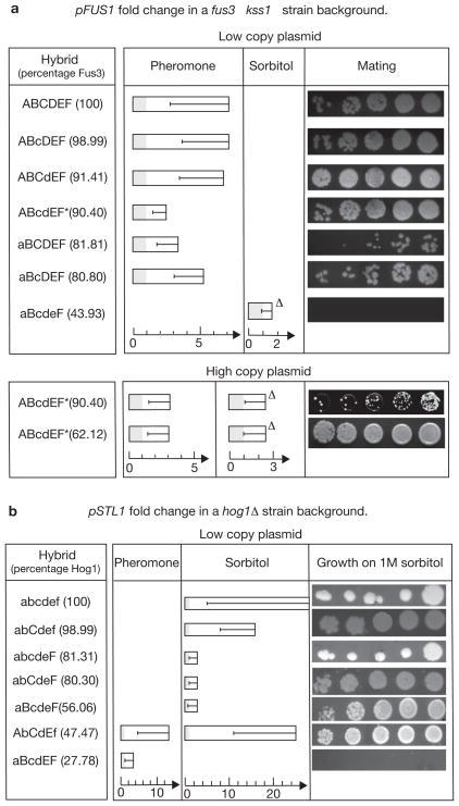
Several hybrid MAPKs function in vivo to faithfully transduce and cross-wire the pheromone and hyper-osmolar signals. (a) The mean fold change in pFUS1 activity (upper panel, measured by a fluorescent reporter) in ~100 induvidual fus3Δ kss1Δ haploid cells containing the hybrid MAPKs expressed from a low-copy plasmid, after stimulation for 2 h by 0.6 μM pheromone or 1 M sorbitol. The percentage of residues in the hybrid that belong to Fus3 and differ from Hog1 is shown in parentheses. Serial dilutions show the mating efficiency of fus3Δ kss1Δ MATa haploids containing the indicated hybrids. Error bars reflect the intrinsically large range of response in the cells and not the measurement of uncertainty in a single cell. One hybrid expressed in low-copy plasmid (upper panel), and two hybrids expressed in high-copy plasmid (lower panel) mediate a cross-wiring whereby sorbitol evokes pFUS1 activity. The persistence of this cross-wiring after deleting STE7 is indicated by ‘Δ’. Hybrids showing constitutive pFUS1 activity are marked with an asterisk. Sorbitol-induced pFUS1 activity of aBcdeF was measured in fus3Δkss1Δhog1Δ cells. (b) The mean fold change in pSTL1 activity measuring an osmolar response, in ~100 single hog1Δ haploid cells containing the hybrid MAPKs and stimulated in the same manner as in a. The axes for fold change are scaled differently for the pheromone and sorbitol stimuli. Serial dilutions show the efficient growth of hog1Δ strains carrying the hybrid on plates containing 1 M sorbitol. Two hybrids mediated a cross-wiring showing pSTL1 activity on pheromone exposure (Supplementary Information, Data Analysis). The pheromone induced pSTL1 activity of aBcdEF was measured in fus3Δ kss1Δ cells.

