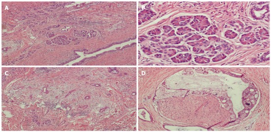
Figure 2.

Histopathology shows the heterotopic pancreas and ductal adenocarcinoma arising from heterotopic pancreas. A: Histopathology showing the pancreatic tissue consisting of acinar cells and ductal elements [hematoxylin and eosin (HE), × 4]; B: Histopathology showing the acinar cells containing eosinophilic granules (HE, × 10); C: The solid area showing ductal adenocarcinoma arising from heterotopic pancreas with nerve infiltration (HE, × 4); D: The solid area showing ductal adenocarcinoma arising from heterotopic pancreas with nerve infiltration (HE, × 20).
