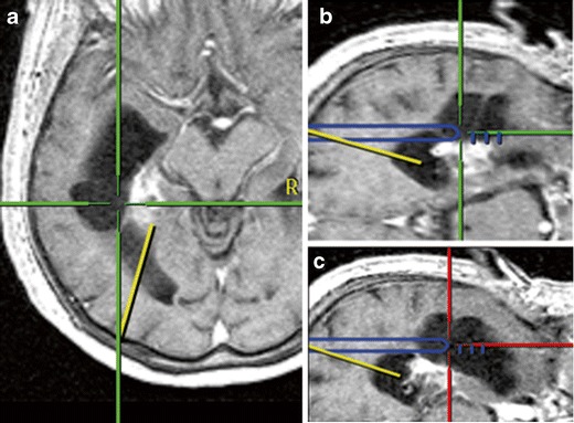
Fig. 4.

A 70-year-old female with a left temporal AVM which was treated radiosurgically. The patient developed an intraventricular cyst which was progressive in volume and caused a visual field defect. During the endoscopic procedure with neuronavigationally tracked endoscope tip, the small occipital horn was first entered following the predefined yellow trajectory (left), then, on a new trajectory, the cyst was entered (middle) and finally opened to the temporal horn (right). Navigation was essential for ventricle puncture and anatomical orientation
