
Figure 5. Immunofluorescence images of Ad-EDNRB transduced and nontransduced oral SCC cells.
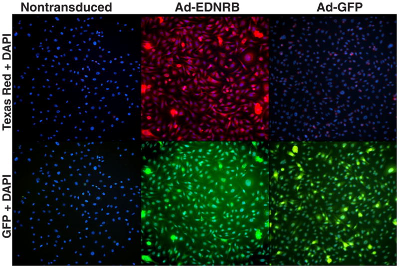
Images are shown at 20X magnification. Images are taken from a single exposure setting. The top row of images shows Texas Red and Hoechst staining. The bottom row of images shows GFP and Hoechst staining. ETB receptor antibody was secondarily tagged with Texas Red and cell nuclei were counterstained with Hoechst. Nontransduced and Ad-GFP transduced HSC-3 cells display minimal ETB receptor expression, whereas Ad-EDNRB transduced HSC-3 cells exhibit strong ETB receptor expression. Ad-GFP transduced HSC-3 cells serve as the positive control for transduction. Ad-GFP transduced HSC-3 cells show strong GFP expression. Ad-EDNRB transduced HSC-3 cells also show GFP expression due to a C-terminal GFP tag on the ETB receptor protein.